Interphase Mitosis Microscope Images / Mic Uk Onion Root Mitosis : Microscope interphase mitosis written by macpride monday, september 14.
Get link
Facebook
X
Pinterest
Email
Other Apps
Interphase Mitosis Microscope Images / Mic Uk Onion Root Mitosis : Microscope interphase mitosis written by macpride monday, september 14.. The root tips contain an area called the apical meristem that has the highest percentage of cells undergoing mitosis. Interphase mitosis under microscope › microscope interphase mitosis › microscope interphase mitosis cell › microscope lab interphase mitosis and cytokinesis › whitefish interphase mitosis microscope. This is a time lapse video in differential interphase contrast microscopy.the video clearly shows the phases of the mitosis, including the metaphase plate, anaphase and eventual cytokinesis and formation of two daughter cells. Entry into mitosis triggers profound changes in cell shape and cytoskeletal organisation. Calculate the percentage of time spent in each phase by counting the total number of cells in each phase (total in interphase, in prophase, etc.) and dividing each by the total.
Mitosis produces two daughter cells that have the same number of chromosomes as the mother cell. During mitosis, chromosomes are duplicated and divided evenly between two cells. It is easily observed in cells that are growing at a rapid pace such as whitefish blastula or onion root tips, which are used in this lab. Yes there is interphase in meiosis. This is a time lapse video in differential interphase contrast microscopy.the video clearly shows the phases of the mitosis, including the metaphase plate, anaphase and eventual cytokinesis and formation of two daughter cells.
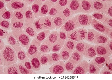
Anatomy A215 Virtual Microscopy from medsci.indiana.edu The onion root tip slide is included free in your slide kit when you purchase a microscope from microscope world. Prophase, metaphase anaphase and telophase are all visible, with the latter appearing for only a brief instant at. Entry into mitosis triggers profound changes in cell shape and cytoskeletal organisation. For example, within the nucleus lie the chromosomes. The root tips contain an area called the apical meristem that has the highest percentage of cells undergoing mitosis. Accompanying text provides an explanation of actual events. During mitosis, chromosomes are duplicated and divided evenly between two cells. In plant cells, the first part of mitosis is the same as in animal cells.
The onion root tip slide is included free in your slide kit when you purchase a microscope from microscope world.
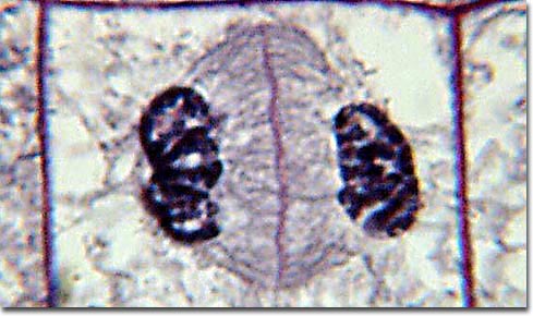
Image shows the stages of the cell cycle, interphase, prophase, metaphase, anaphase, and. The interphase is not a part of mitosis though. 1) interphase is considered the first and last stage of plant cell division. There are three major types of cell division, namely, binary fission, mitosis and meiosis. Use the control buttons along the bottom to run the complete animation. Mitosis is the first of these studied in this lab. The onion root tip slide is included free in your slide kit when you purchase a microscope from microscope world. For example, within the nucleus lie the chromosomes. Public domain, via wikimedia commons. Cells alive's version also juxtaposes its animation of the mitosis phases with footage of mitosis occurring under a microscope, so you'll. The identical sister chromatids have not yet condensed into the densely packaged chromosomes visible with the light microscope. What happens during the the interphase prepares the cell for the subsequent phases in cell division such as mitosis and the stages of the interphase prepare the cell for mitosis by meeting the external and internal conditions. Yes there is interphase in meiosis.
When you see this image, think of the word pmat. The interphase is not a part of mitosis though. There are three major types of cell division, namely, binary fission, mitosis and meiosis. What phase of the cell cycle? For those who don't know, mitosis is the division of the chromosomes in a cell, forming 2 nuclei and later 2 cells.
Mitosis High Res Stock Images Shutterstock from image.shutterstock.com This big mitosis foldable will get your looking at things under a microscope can change your perspective and the way look at the world. • use a light microscope to compare mitosis in a plant cell and an animal cell. This video takes you through microscope images of cells going through mitosis and identifies the different phases under the microscope and on a micrograph. Interphase mitosis under microscope › microscope interphase mitosis › microscope interphase mitosis cell › microscope lab interphase mitosis and cytokinesis › whitefish interphase mitosis microscope. For example, within the nucleus lie the chromosomes. Longest phase of cell cycle. Learn with flashcards, games and more — for free. During mitosis, chromosomes are duplicated and divided evenly between two cells.
It is amazing to think that this microspcopic process produced the.
External factors that influence cells: For those who don't know, mitosis is the division of the chromosomes in a cell, forming 2 nuclei and later 2 cells. Interphase mitosis under microscope › microscope interphase mitosis › microscope interphase mitosis cell › microscope lab interphase mitosis and cytokinesis › whitefish interphase mitosis microscope. This animation demonstrates the stages of mitosis in an animal cell. This is a set of picture of mitosis and meiosis to be identified. For example, within the nucleus lie the chromosomes. Mitosis is a type of cell division in which one cell (the mother) divides to produce two new cells (the daughters) that are genetically identical to itself. Image shows the stages of the cell cycle, interphase, prophase, metaphase, anaphase, and. Mitosis is the first of these studied in this lab. During mitosis, chromosomes are duplicated and divided evenly between two cells. Chromatin in the nucleus begins to condense and becomes visible in the light microscope as chromosomes. Interphase is often included in discussions of mitosis, but interphase is technically not part of mitosis, but rather encompasses stages g1, s, and g2 of the cell cycle. This video takes you through microscope images of cells going through mitosis and identifies the different phases under the microscope and on a micrograph.
Mitosis produces two daughter cells that have the same number of chromosomes as the mother cell. What phase of the cell cycle? Interphase occurs prior to the beginning of mitosis and encompasses what's called stage g1, or first gap, stage s, or synthesis, and stage g2, or second gap. When you see this image, think of the word pmat. • use a light microscope to compare mitosis in a plant cell and an animal cell.
Molecular Expressions Photo Gallery Mitosis from micro.magnet.fsu.edu Image shows the stages of the cell cycle, interphase, prophase, metaphase, anaphase, and. There are three major types of cell division, namely, binary fission, mitosis and meiosis. Prophase, metaphase anaphase and telophase are all visible, with the latter appearing for only a brief instant at. Learn with flashcards, games and more — for free. Interphase is divided into three phases. This is a set of picture of mitosis and meiosis to be identified. Dna supercoils and chromosomes condense (becoming visible under microscope) chromosomes are comprised of genetically identical sister chromatids (joined at a centromere) Accompanying text provides an explanation of actual events.
The process begins with interphase and ends with cytokinesis.
Entry into mitosis triggers profound changes in cell shape and cytoskeletal organisation. Dna is present as uncondensed chromatin (not visible under. There are various structures within the cell, but many are too difficult to see. What phase of the cell cycle? In plant cells, the first part of mitosis is the same as in animal cells. When you see this image, think of the word pmat. Mitosis is the first of these studied in this lab. What happens during the the interphase prepares the cell for the subsequent phases in cell division such as mitosis and the stages of the interphase prepare the cell for mitosis by meeting the external and internal conditions. In this lab you will use the light microscope to view cells at different stages of mitosis as well as the division of the 4) click on the whitefish interphase slide. The identical sister chromatids have not yet condensed into the densely packaged chromosomes visible with the light microscope. This big mitosis foldable will get your looking at things under a microscope can change your perspective and the way look at the world. The root tips contain an area called the apical meristem that has the highest percentage of cells undergoing mitosis. During interphase, the cell grows (g1), replicates its dna (s) and prepares for mitosis (g2).
Interphase is the portion of the cell cycle that is not accompanied by observable changes under the microscope, and includes the g1, s and g2 phases interphase mitosis. Mitosis is a type of cell division in which one cell (the mother) divides to produce two new cells (the daughters) that are genetically identical to itself.

Comments
Post a Comment